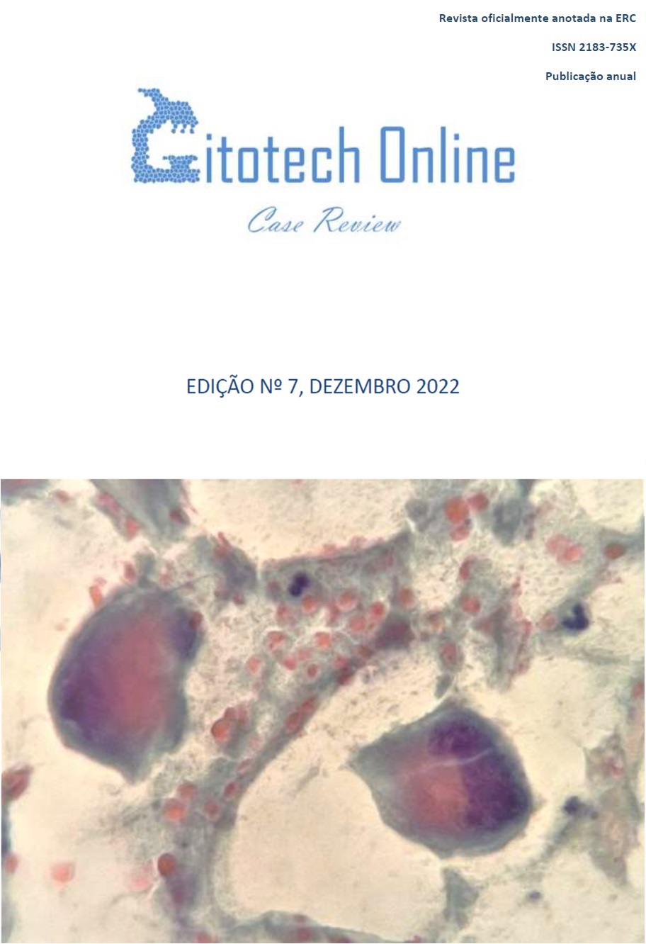
Cytology applied to the animal field: fibroadenoma in female cat, discussion of a clinical case
DOI:
https://doi.org/10.26537/citotech.vi7.4926Keywords:
companion animals, breast tumours, cat, Fibroadenoma, Phyllodes tumorAbstract
Biological, genetic, histopathological, molecular, and morphological similarities have been made between human tumours and spontaneous neoplasms in pets. As such, spontaneous neoplasms in companion animals have been recognized as advantageous for the study of comparative oncobiology. Although it is difficult to estimate their prevalence and incidence, it is known that breast tumours are the most frequent type of neoplasm found in female dogs and the third most frequently observed in female
cats. This article presents a clinical case of a mammary nodule from an 8-year-old female cat, whose cytological smear, stained by Giemsa's method, revealed the presence of sets of ductal cells with monolayer distribution and slight atypia, rare myoepithelial cells, macrophages, and background with some associated serosity. The purpose of this paper is to share information regarding the animal field,
highlighting the transversal nature of cytology with the presentation of a clinical case and correlation between its cytology and histology. The cytological result attributed was of a fibroadenoma, however, its histology revealed to be a phyllodes tumor.
Downloads
Published
How to Cite
Issue
Section
License

This work is licensed under a Creative Commons Attribution-NonCommercial-NoDerivatives 4.0 International License.


